
Myositis Ossificans
Characteristics
– Inflammatory pseudotumor
– Muscle lesion (most frequently in long muscles of the extremities)
– Heterotopic calcifications
– Relatively well-defined contour
– Inhomogeneous appearance
Predisposing Factors
– History of trauma
– Paralysis
– Burns
– Tetanus
– Intramuscular hematoma
– Hereditary disorders (fibrodysplasia ossificans progressiva)
Clinical Presentation
– Rapidly growing soft tissue mass
– Muscle pain
Phases / Lesion Development
– Muscle injury with peripheral and centripetal calcification
– Acute phase: 1–7 days, myxoid matrix
– Subacute phase: 7–15 days, osteoid matrix
– Late phase: more than 15 days, mature bone in «layers»
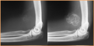
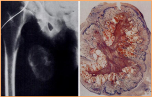
X-Ray Findings
– Late visualization (mature bone)
– Heterotopic soft tissue calcification
– Distant from the adjacent bone
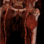
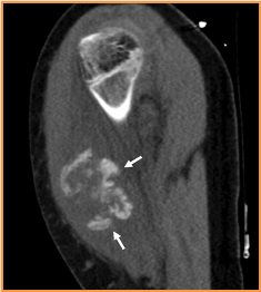
CT Findings
– Detects calcifications earlier than plain X-ray
– Heterotopic calcification
– Hypodense center due to adipose metaplasia
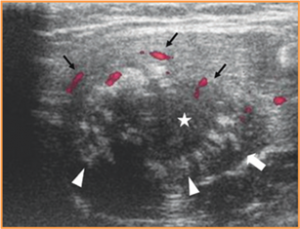
Ultrasound Findings
Demonstrates the «zonal» phenomenon:
– Peripheral hypoechoic zone (hyperemia)
– Middle hyperechoic zone (calcifications)
– Central hypoechoic zone (fibroblastic stroma)
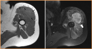
MRI Findings
– Intramuscular, circumscribed lesion
– May show the «zonal» phenomenon
– T1–T2: iso/hyperintense, inhomogeneous
– Areas of low signal (calcium)
– Contrast: ring enhancement in acute and late lesions; diffuse/heterogeneous enhancement
– Adjacent muscle fibers are continuous
– In the intermediate phase may show areas of fibrous appearance (hypointense) resembling nodular fasciitis
– Edema of adjacent muscle fibers
Definitive Diagnosis – Pathological Anatomy
– Time is a diagnostic factor
– Biopsy when imaging is inconclusive or atypical
– Differentiation from malignant lesions
– Histology: cytology is inconclusive in acute and subacute phases; definitive in late lesion with the «zonal» phenomenon; biopsy core must span the full thickness of the lesion
Differential Diagnoses
– Soft tissue sarcoma (malignant fibrous histiocytoma, osteosarcoma)
– Proliferative myositis
– Traumatic lesion
– Sarcoidosis
– Infectious-inflammatory process
– Nodular fasciitis
– Metastases
Conclusion
– Frequent history of trauma
– «Zonal» pattern
– Imaging: MRI, ultrasound, CT, X-ray
– Temporal evolution
– Biopsy to confirm or rule out other diagnoses
Bibliography
Myositis Ossificans: MR Appearance with Radiologic-Pathologic Correlation, AJR.
Case 118: Proliferative Myositis, Radiology: Volume 244: Number 2—August 2007
For any comments or suggestions: radiologyzones@gmail.com
This material was automatically translated from medicosradiologos.com.ar