Clinical Case: Osteoid Osteoma
Case Presentation
A 23-year-old male patient presents with pain in the hallux (big toe) with 2 months of evolution. The pain is predominantly nocturnal, and there is no significant history of trauma.
Imaging Findings
X-Ray and Computed Tomography
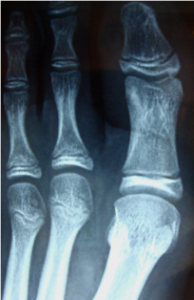
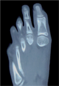
In the proximal phalanx of the hallux, on the lateral border of the distal epiphysis, an ovoid image of greater density than the adjacent bone is observed.
Does anything stand out in these images?
If we look closely, on the proximal phalanx of the hallux, above the lateral border of the distal epiphysis, we can see an ovoid shape, denser than the adjacent bone.
Differential Diagnoses Considered
– Enostosis / Bone island
– Osteoid osteoma
– Osteoblastoma
– Cortical thickening due to infectious process
– Intracortical hemangioma
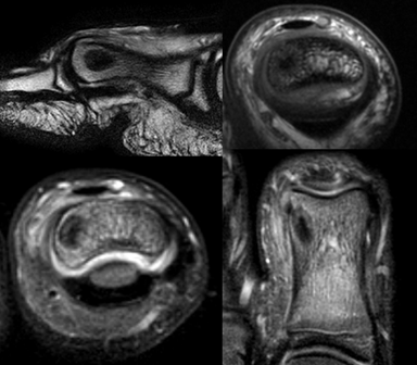
MRI performed with a microcoil, demonstrating a low signal intensity lesion with a more hypointense center, along with edema of adjacent bone trabeculae, increased intra-articular fluid, and soft tissue edema.

Definitive Diagnosis
The clinical presentation was key: nocturnal pain relieved by NSAIDs confirmed the diagnosis of osteoid osteoma. Nuclear medicine imaging showed a focus of hypercaptation in the right hallux, corroborating the diagnosis.
![]()
Clinical Conclusions
High-resolution multislice CT with a small field of view and targeted reconstructions allows visualization of the characteristic «nidus» without the need for additional studies.
You can read more about Osteoid Osteoma in the following article:
Radiologic Diagnosis of Osteoid Osteoma: From Simple to Challenging Findings.
For any comments or suggestions: radiologyzones@gmail.com
This material was automatically translated from medicosradiologos.com.ar