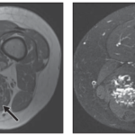

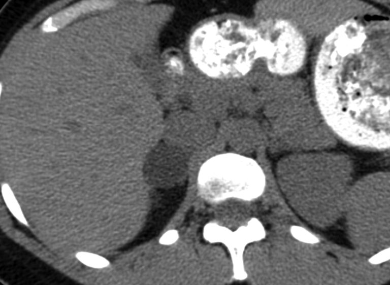
Increasingly, radiologists are encountering an unexpected finding: a small adrenal mass discovered incidentally during a computed tomography (CT) scan ordered for another reason. This is estimated to occur in approximately 4 to 5% of all abdominopelvic CT scans, and its prevalence increases with age. Consequently, the term adrenal incidentaloma has become increasingly common in everyday radiological practice.
The vast majority of these lesions are benign. In patients with no history of malignancy, the most frequent diagnosis is adrenal adenoma, which accounts for approximately 75% of incidental findings. However, the possibility—although statistically small—that an adrenal mass may correspond to a metastasis, adrenocortical carcinoma, or pheochromocytoma requires the radiologist to have a precise understanding of the diagnostic criteria that allow for reliable characterization and avoidance of unnecessary procedures.
In this summary article, we offer an overview of the topic: from the anatomy and physiology of the adrenal glands to the historical evolution of imaging methods used to study adrenal adenomas, with particular attention to the advances of recent decades. The reader will find here not only the classic foundations—densitometry on non-contrast CT and the calculation of washout—but also the most recent discussions that challenge paradigms held for more than twenty years, and emerging technologies such as dual-energy CT, which promise to simplify the diagnostic algorithm without sacrificing accuracy.
Anatomy and function of the adrenal glands
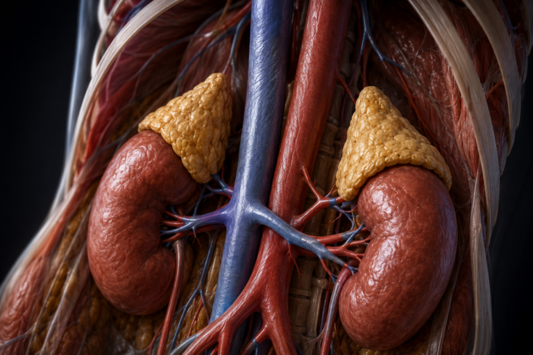
The adrenal glands are paired retroperitoneal organs located in the perirenal space, adjacent to the upper pole of each kidney, and surrounded by adipose tissue. They weigh between 4 and 6 grams each and measure approximately 3 to 5 centimeters along their longest axis. Despite their small size, their blood supply is exceptionally rich: they receive branches from the inferior phrenic artery, directly from the aorta, and from the renal artery, making them one of the organs with the highest blood flow per gram of tissue in the human body.
The shape differs between the two sides, a relevant detail for the radiologist: the right pancreatic gland is typically pyramidal or triangular, nestled between the upper pole of the right kidney, the right hepatic lobe, and the inferior vena cava; the left has a semilunar or crescent shape, with a larger surface area and is closer to the body of the pancreas, the splenic artery, and the stomach. This morphological asymmetry is normal and should not be interpreted as pathological in imaging studies.
From a functional standpoint, the adrenal gland is actually two embryologically and functionally distinct organs fused into a single structure. The adrenal cortex, of mesodermal origin, constitutes approximately 80–90% of the glandular volume and is organized into three well-differentiated concentric zones. The zona glomerulosa, the outermost zone, is the sole producer of aldosterone, the mineralocorticoid that regulates sodium and potassium balance and, consequently, blood pressure, under the control of the renin-angiotensin system. The zona fasciculata, the largest zone, synthesizes cortisol, the major glucocorticoid, whose secretion is stimulated by pituitary ACTH and regulates carbohydrate, protein, and lipid metabolism, the immune response, and the stress response. The reticular zone, the innermost part of the cortex, produces adrenal androgens—mainly DHEA and androstenedione—which play a relevant role in pubertal development and in the regulation of the sexual axis.
The adrenal medulla, of ectodermal origin (derived from the neural crest), occupies the center of the gland and can be considered a specialized sympathetic ganglion. Its chromaffin cells secrete catecholamines—mostly adrenaline and noradrenaline—in response to stimuli from the autonomic nervous system, mediating the fight-or-flight response.
This dual embryological nature directly explains the spectrum of tumors that arise in the gland. Adenomas —the most frequent finding in routine radiological practice—are benign neoplasms of the cortex, and their most characteristic diagnostic feature is precisely their intracellular lipid content, reflecting the large reserves of cholesterol that cortical cells accumulate as a hormonal precursor. This intracellular fat is the pathophysiological basis of all the densitometric criteria that will be analyzed in the following sections. Pheochromocytomas , on the other hand, are tumors of the medulla, without significant lipid content and with a completely different dynamic behavior on contrast-enhanced computed tomography. This different architecture is related to the foundation of adrenal radiological semiotics.
Methods for studying adrenal adenoma: evolution and diagnostic criteria
The first major advance in the radiological characterization of adrenal adenomas was conceptually simple: cortical adenomas accumulate large amounts of intracellular lipids—mainly cholesterol and its esters, raw materials for hormone synthesis—and this fat reduces tissue density as measured in Hounsfield units (HU) on non-contrast CT. Malignant lesions and metastases, on the other hand, have low lipid content and present with higher densities.
The 10 HU threshold was established as the gold standard following a meta-analysis of multiple studies published in the late 1990s: an adrenal mass with a density of 10 HU or less on non-contrast CT has a sensitivity of 71% and a specificity of 98% for the diagnosis of adenoma. High specificity means that when the criterion is met, the diagnosis is almost certain; the limitation lies in the sensitivity: up to 30% of adenomas are lipid-poor and exceed this threshold, requiring further evaluation.
For lipid-poor adenomas—those with a baseline density greater than 10 HU—the solution came with the observation that cortical adenomas wash out iodinated contrast medium much more rapidly than malignant lesions. This phenomenon is due to differences in vascularization and the characteristics of the tumor interstitium. Adenomas have fenestrated capillaries with rapid washout; metastases and carcinomas retain contrast longer in larger interstitial spaces.
The CT washout protocol consists of three phases: baseline without contrast, portal phase at 60–70 seconds, and delayed phase at 15 minutes. Two indices are calculated from these three values: absolute washout ((portal HU − delayed HU) / (portal HU − baseline HU) × 100)) and relative washout ((portal HU − delayed HU) / portal HU × 100)). Relative washout is used when a baseline phase is unavailable. An absolute washout value greater than or equal to 60% or a relative washout value greater than or equal to 40% is considered diagnostic.
The work of Korobkin et al. (1998) demonstrated that at 15 minutes post-contrast, adenomas and non-adenomas could be differentiated with 96% sensitivity and specificity using an attenuation threshold of 37 HU in the delayed phase. The study by Caoili et al. (2002) combined both strategies—baseline and washout—in a unified protocol that achieved 98% sensitivity and 92% specificity in 166 masses. This protocol became the standard and was incorporated into the ACR guidelines in 2017.
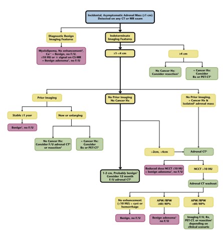
The 2017 American College of Radiology guidelines (Mayo-Smith et al.) systematized all this evidence into a decision algorithm. The entry point is baseline density: if the mass measures ≤10 HU, it is a lipid-rich adenoma and requires no further investigation. If it exceeds this threshold, the washout protocol is indicated; an APW ≥60% or RPW ≥40% confirms an adenoma. Size is also integrated into the algorithm: masses >4 cm have a higher probability of malignancy and warrant more proactive management regardless of densitometric values.
The Achilles’ heel of the classic protocol is the need for three phases, with the resulting additional radiation dose and logistical complexity. Dual-energy computed tomography (DECT) offers a promising alternative by characterizing tissues through simultaneous acquisition at two energy levels, allowing the calculation of virtual non-contrast attenuation (VNC), tissue fat fraction, and iodine density in a single portal phase.
The study by Loonis et al. (2023) on 139 lesions demonstrated that a fat fraction ≥23.8% achieves 59% sensitivity with 100% specificity for adenoma, surpassing VNC attenuation. The combination of a fat fraction ≥23.8% or a relative enhancement ratio ≥214% increases sensitivity to 68% while maintaining 100% specificity, all without the need for a baseline or delayed phase.
Differential diagnoses of adrenal adenoma
Adrenal adenoma is the most common adrenal lesion, but it is not the only one. Radiologists evaluating adrenal masses must be aware of a spectrum of alternative diagnoses that may morphologically overlap with adenoma, especially when the lesion is lipid-poor or has atypical features. The diagnostic key—as in all radiology—lies in integrating imaging findings with the clinical context and biochemical data.
Among all the differential diagnoses, pheochromocytoma deserves special mention because it has the most serious consequences if overlooked. It can present with variable densities, intense and heterogeneous enhancement, and—as demonstrated by multiple studies, including a 2018 meta-analysis—up to 47% meet washout criteria for adenoma when the classic protocol is applied. In lesions smaller than 3 cm, this rate can reach 80%. The practical consequence is clear: any adrenal mass with a density greater than 10 HU requires determination of metanephrines in plasma or fractionated urine before any biopsy or surgical procedure, regardless of the washout result.
Adrenal corticotropin-releasing carcinoma ( ACC) is rare—with an incidence of 1–2 cases per million inhabitants per year—but carries a poor prognosis when diagnosed late. Its main alarming feature on imaging is its size: ACR guidelines recommend considering direct surgical resection without prior biopsy for any mass larger than 4 cm without a history of known extra-adrenal malignancy. Morphologically, it is usually large, heterogeneous, with areas of necrosis and calcifications, and may extend into the vena cava.
In patients with known extra-adrenal neoplasia, the pretest probability of adrenal malignancy changes dramatically. However, even in this scenario, most adrenal masses are benign adenomas. The key point is that in this context, washout and densitometry remain useful, but they should be complemented with PET-CT when doubt persists, given that adrenal metastases tend to be FDG-avid, with higher uptake than the liver. It is worth emphasizing that truly isolated and incidental metastases in patients without other disseminated disease are extremely rare.
Conclusion
In the radiologist’s daily practice, the adrenal adenoma is a finding that one lives with: it appears unintentionally, in studies ordered for reasons completely unrelated to the gland, and requires a precise diagnostic response that avoids both the over-investigation of benign lesions and the unnoticed passage of a malignant or functioning pathology.
This article has attempted to trace the evolution of this response over more than four decades. It began with the observation that cortical adenomas accumulate intracellular lipids, and that this fat translates into low densities on non-contrast CT scans—the fundamental finding that gave rise to all subsequent diagnostic criteria. Then came the understanding of the dynamic behavior of contrast: adenomas wash out quickly, malignant lesions retain it. This observation led to the washout protocol that dominated practice for more than twenty years and was formalized by the ACR guidelines in 2017.
In recent years this washout protocol has been questioned and other ways of assessing adrenal lesions and diagnosing adenomas have been proposed.
Below you will find the corresponding bibliography ordered according to the publication years of the selected articles, from the oldest to the most recent.
Literature:
CT Time-Attenuation Washout Curves ofAdrenal Adenomas and Nonadenoma (1998)
Adrenal Masses: Characterization with Combined Unenhanced and Delayed Enhanced CT (2002)
Management and diagnosis of adrenal incidentaloma (2011)
Washed up: the end of an era for adrenal incidentaloma CT (2023)
This material was automatically translated from medicosradiologos.com.ar
For any comments or suggestions: radiologyzones@gmail.com