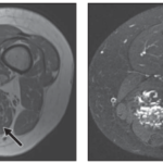

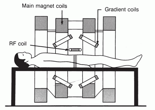
In order to perform the best possible quality magnetic resonance imaging studies, as well as in any of the other diagnostic imaging techniques, it is necessary to know the basic concepts of the method, the contrast media and the proper positioning of the patient in the equipment, as well as the correct cutting plane of each region, even more so with regard to the osteoarticular system.
While it has long been believed that magnetic resonance imaging (MRI) is not an operator-dependent method, our experience shows that it is. Two studies from equipment with the same field of view can have very different qualities depending on the processing of the sequences, their selection, the use of appropriate coils, and the slice planes employed.
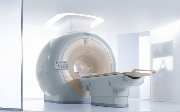
The more information we have about the method we are using and the equipment available for the study, the better the result will be. Similarly, the more we know about the anatomical region and the pathology being studied, the greater the likelihood of arriving at a more accurate diagnosis. The collaborative work of medical specialists and imaging technicians is what will produce a high-quality study.
We offer two essential texts to help you understand magnetic resonance imaging (MRI). The second text also includes a guide to the imaging planes used for studying joints.
Magnetic Resonance Imaging. Part 1: the Basic Principles
Basic Principles and Terminology of Magnetic Resonance Imaging
This material was automatically translated from medicosradiologos.com.ar
For any comments or suggestions: radiologyzones@gmail.com