
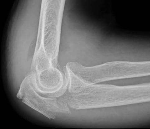
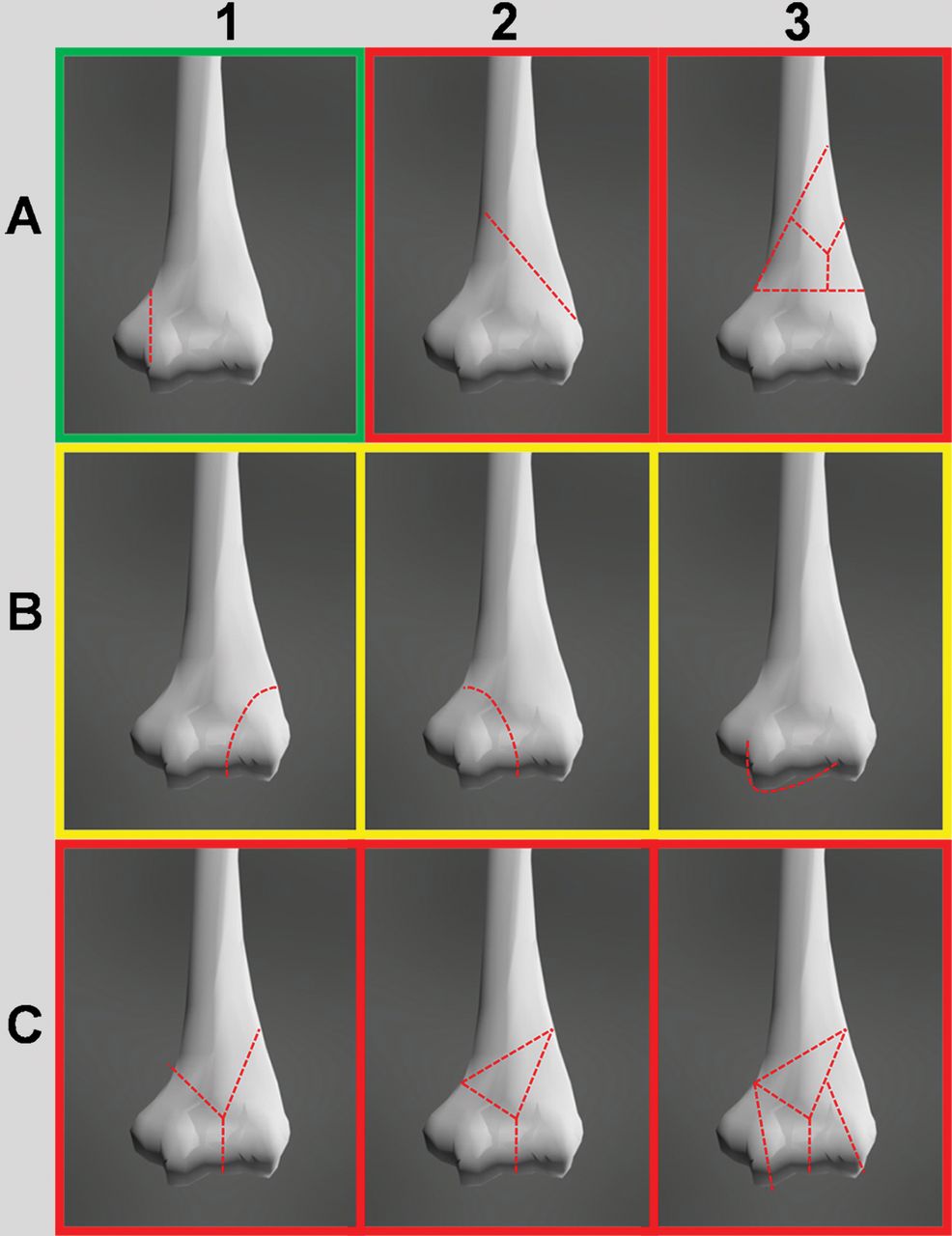
The elbow joint is composed of several articulations (humeroradial, humeroulnar, proximal radioulnar), which adds complexity to the analysis of traumatic elbow injuries. As we always mention, the essential element when evaluating any joint or any anatomical region is a thorough knowledge of anatomy, for which both classic texts (here we cite some examples) and iconographic atlases such as Netter ‘s are useful .
Once the anatomy is understood, including the essential location of the various ligaments, neural structures, and blood vessels, we must determine how the injury occurred, that is, the mechanism of injury. This can be easily determined by questioning the patient and inspecting the joint for secondary signs of injury such as soft tissue swelling, bruising, and other signs. It is also helpful to be familiar with certain joint assessment maneuvers, which can help anticipate the injury.
Once the mechanism of injury is known, a diagnostic suspicion must be formulated. We must hypothesize what the patient has injured: fractures, ligamentous injuries, nerve damage, etc. While this task falls to the orthopedic surgeon, it will be very helpful for us to perform it and then confirm it with them. It will be much easier to examine the patient if we know what we are looking for, allowing us to direct the examination and focus on finding the suspected injury. This will also help us decide what type of study we need to perform, whether it be X-rays, CT scans, MRI, or ultrasound. In this way, we will also avoid performing unnecessary tests.
Here is a useful article for evaluating elbow injuries:
Traumatic Elbow Injuries: What the Orthopedic Surgeon Wants to Know
This material was automatically translated from medicosradiologos.com.ar
For any comments or suggestions: radiologyzones@gmail.com