
The objective of presenting the following case is to propose a systematic approach for studying soft tissue injuries, given that their diagnosis is often extremely complex. It is important to note that imaging alone is often insufficient to identify the specific injury. Therefore, the key to writing the report is to describe the characteristics of the injury, taking into account the following parameters:
- Location : whether the lesion is located in the cutaneous plane, in the subcutaneous cellular tissue, or in the muscular plane.
- Extent : whether the injury extends to adjacent structures or planes. In the case of a very extensive injury, determining the point of origin can be complex. Always look for involvement of adjacent bone planes.
- Size : Measure the lesion in all three planes, preferably.
- Relationship with neurovascular bundles : describe whether or not they have a cleavage plane with said bundles, whether they displace them, etc.
- Determining the lineage : certain characteristics of the lesion, such as the content of adipose tissue, continuity with neural structures or tendon sheaths, can give us an idea of the lineage of origin of the lesion: adipose, neural, etc.
- Possibility of anatomopathological evaluation : indicate if the lesion is amenable to biopsy and, if so, where the greatest benefit would be obtained. For example, in the case of a lesion with areas of necrosis, we must indicate where or which are the solid areas to be biopsied.
If we can describe all these characteristics in the report, we will be making a great contribution to the diagnosis, even if we cannot give the injury a «name and surname,» which is not always possible and can even lead to unnecessary errors.
In summary, our role as radiologists is to accurately describe, attempt to characterize the nature of the lesion, and propose differential diagnoses.
A male patient, 84 years old, presents to the consultation at our diagnostic center, who consults for a palpable, slow-growing mass in the root of the left thigh.
As a first study, a simple x-ray of the left thigh is performed, which we show below.
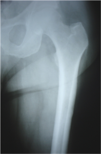
In this radiograph, due to the folds of adipose tissue and the overlapping layers, no lesions can be identified with the naked eye. The bone structure shows no abnormalities.
The magnetic resonance imaging study for which the patient came was then performed, obtaining these images:
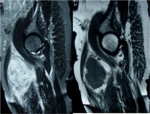
These images correspond to sagittal STIR and T1-weighted sections. A soft tissue mass with lobulated contours and heterogeneous signal is identified, consistent with a solid lesion, which does not appear to have cystic or necrotic areas. It is associated with edema of the adjacent soft tissues, which it displaces, but does not affect the neighboring bone.
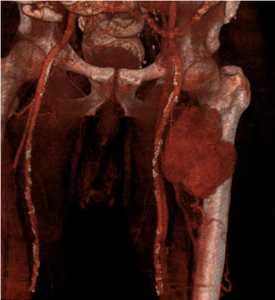
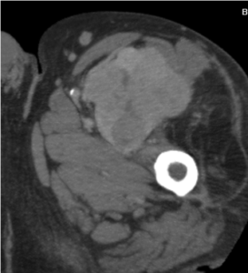
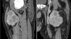
A computed tomography angiography with three-dimensional reconstruction was also performed to evaluate its relationship with vascular structures, obtaining these images:
Differential diagnoses initially involve considering the type of tumor: neural, adipose, vascular.
The CT scans show areas of adipose density in the lower portion of the lesion, surrounding the solid lesion. This is suggestive of an adipose lesion. The solid areas exhibit heterogeneous density, with areas of necrosis and a multilobulated appearance. The femoral vascular bundle is displaced, with a clear cleavage plane visible.
We proposed a diagnosis of malignant adipose lesion, which was confirmed in the post-surgical anatomopathological study as LIPOSARCOMA .
We’ve included two articles you can read on the subject, from Radiographics and Radiology:
2. Liposarcoma
For any comments or suggestions: radiologyzones@gmail.com
This material was automatically translated from medicosradiologos.com.ar