
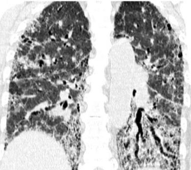
The pathology of the pulmonary interstitium, and within it, interstitial pneumonias result in a complex diagnostic challenge, in which, often, a final result is not obtained until a lung biopsy has been performed.
High-resolution computed tomography (HRCT) has made significant contributions to the study of interstitial pneumonias, as it allows for the assessment of lung involvement and helps in diagnosing various conditions. It is clear that among the numerous possibilities, arriving at the correct diagnosis is often extremely complex; we must remember that the radiologist must always narrow down the long list of differential diagnoses to the fewest possible options. Dialogue with the patient and the pulmonologist will always provide valuable information for the study.
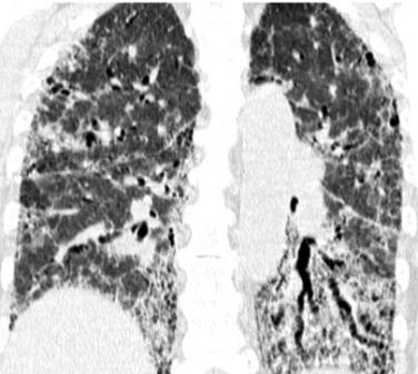
Each entity must carry out its image acquisition protocols to minimize the margin of error, and the protocols must be respected and updated according to the advances in each area.
We are sharing two articles published in 2003 and 2007, which, while they require updating, provide essential information for diagnosis, particularly by bringing order to such a broad pathology. We are confident that by considering the general guidelines in these articles, the approach to this pathology will be less complex.
- CT-Histologic Correlation of the ATS/ERS 2002 Classification of Idiopathic Interstitial Pneumonias
- What Every Radiologist Should Know about Idiopathic Interstitial Pneumonia
This material was automatically translated from medicosradiologos.com.ar
For any comments or suggestions: radiologyzones@gmail.com