
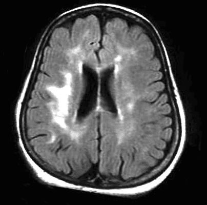
White matter lesions are a common finding in brain imaging and have various causes. Depending on their characteristics, they are representative of different types of pathologies. In 1987, Dr. Fazekas conducted a study and published an article describing and classifying these lesions, thus creating the Fazekas scale for white matter lesions.
White matter lesions, frequently observed on MRI scans of older adults, are associated with various geriatric disorders, including cerebrovascular and cardiovascular diseases, dementia, and psychiatric disorders. Currently, white matter lesions are divided into periventricular and deep lesions. While the meaning of these terms varies across studies and this dichotomy remains under debate, a potential difference in pathogenic mechanisms between periventricular and deep lesions provides some clues to understanding the pathophysiology of many geriatric syndromes associated with these lesions.
The Fazekas scale is simply used to quantify the amount of T2 hyperintense lesions of the white matter that are generally attributed to chronic small vessel ischemia, although, evidently, not all of these lesions are due to this.
This classification was proposed by Fazekas et al. in 1987 and remains a widely used system for describing the severity of white matter diseases in the literature. It is also frequently used in clinical practice, although terms such as «mild,» «moderate,» and «severe» are generally preferred.
The scale is named after Franz Fazekas (born in 1956), an Austrian neurologist and author of the original article published jointly in 1987 in the American Journal of Roentgenology and the American Journal of Neuroradiology.
Classification
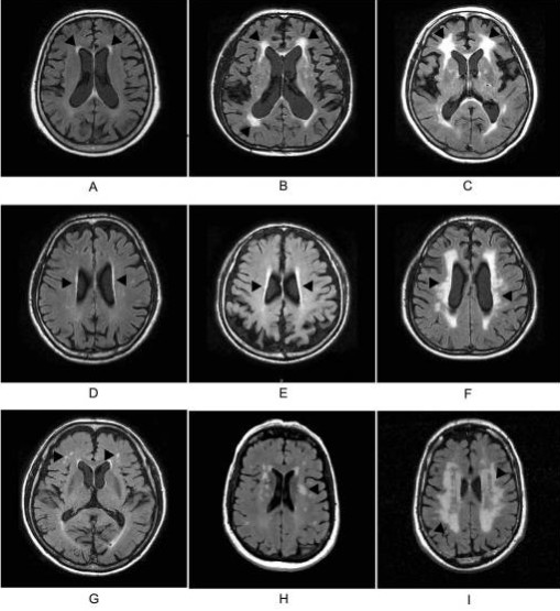
The scale divides the white matter into periventricular and deep white matter, and each region is assigned a grade depending on the size and confluence of the lesions 1.
Periventricular white matter (PVWM)
0 = absent
1 = “caps” or thin coating like a pencil
2 = soft “halo”
3 = irregular periventricular signal extending into the deep white matter
Deep white matter (DBM)
0 = absent
1 = point foci
2 = beginning of the confluence
3 = large confluent areas
We share with you the original article on the Fazecas classification and a general article on white matter lesions.
MR Signal Abnormalities at1.5 T in Alzheimer’s Dementia and Normal Aging
Classification of white matter lesions on magnetic resonance imaging in the elderly
This material was automatically translated from medicosradiologos.com.ar
For any comments or suggestions: radiologyzones@gmail.com