
Diagnostic imaging has become an essential tool for diagnosing various diseases over the last few decades. So much so, that for many pathologies, precise diagnostic and staging systems based on imaging have been developed, which are used in daily medical practice to establish treatments, monitor patients, and make prognoses.
The best-known of these systems is BI-RADS for breast pathology, for which we recently published an update. But, as we mentioned, new systems have emerged over time, such as the one used for prostate cancer, among others.
In general, these are diseases with a high incidence in the population, and their early diagnosis and accurate treatment can significantly improve patients’ health and survival. Specific treatments have also been developed for these diseases, allowing for effective resolution when diagnosed early. This underscores the importance of standardizing diagnostic criteria, so that studies conducted similarly or using standardized protocols can yield comparable results worldwide, thus enabling the establishment of standardized treatments.
Currently, a standardization process is underway for the diagnosis and treatment of pulmonary embolism, and we want to inform you about it.
Pulmonary embolism (PE), defined as the obstruction of the pulmonary arteries, is a common and serious condition that requires accurate diagnosis and standardized reporting. Non-invasive imaging techniques are well-established for the diagnosis and management of PE and are incorporated into multiple scientific society guidelines. However, specific technical recommendations for the optimal interpretation and standardized reporting of computed tomography (CT) angiography and magnetic resonance imaging (MRI) are still lacking. Diagnostic imaging techniques are used to identify the presence or absence of PE and to stratify patient risk. Once an acute clot is identified, the patient’s condition is defined, and the patient is referred for appropriate treatment in a timely manner, the constantly evolving treatment options can be considered. PE management often involves multiple care teams, and clear and consistent communication among them is critical to patient care.
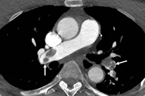
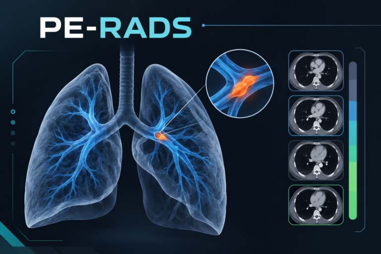
The goal of the Pulmonary Embolism Notification and Data System (PE-RADS) is twofold: firstly, to create a standard lexicon for describing the terms used for the diagnosis of acute PE in CT and MRI angiography, and secondly, to develop a hierarchical and structured reporting system that incorporates anatomical and accessory findings to classify clot location and right heart imaging characteristics in acute PE and to help determine the patient’s cardiovascular status to inform subsequent management.
Essential elements:
1- The Pulmonary Embolism Notification and Data System (PE-RADS) creates a standard lexicon for the description of terms used for the diagnosis of acute pulmonary embolism (PE) in CT and MRI angiography.
2 -PE-RADS provides a structured, hierarchical reporting system that incorporates anatomical and accessory findings to classify clot location and right heart imaging characteristics in acute PE and to help determine cardiovascular status to inform subsequent management.
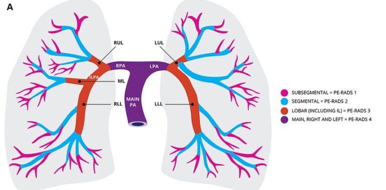
We’re sharing some articles with you to keep you up-to-date on this new diagnostic standardization. The article was published, with minor differences, in Radiology and Chest.
Pulmonary Embolism Reporting and Data System (PE-RADS)
Note: The images were taken from the cited articles.
This material was automatically translated from medicosradiologos.com.ar
For any comments or suggestions: radiologyzones@gmail.com