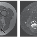

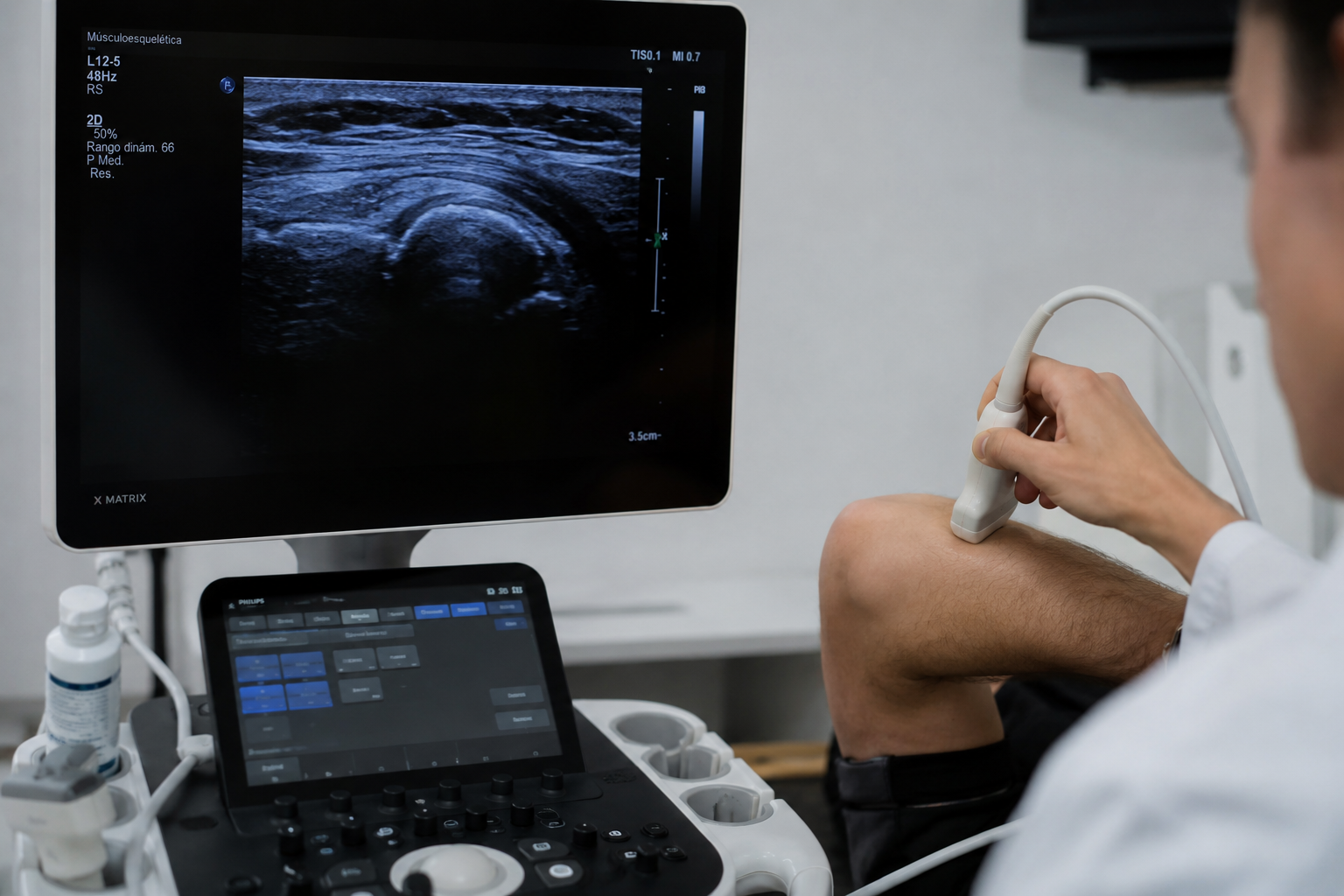
Ultrasound studies of the musculoskeletal system present a certain complexity in their practical execution and in the accuracy of diagnoses. This complexity is often fueled by a certain apprehension among radiologists regarding musculoskeletal pathology, an apprehension we must overcome. For anything we want to do and learn, we first need anatomical knowledge, then good textbooks to begin our studies, knowing where to delve deeper and how to systematize our work so as not to overlook anything. The purpose of this article is to suggest some textbooks and working protocols that will facilitate radiologists’ approach to this area. Furthermore, each radiologist must be familiar with the equipment (ultrasound machine) they work with, its limitations, and its tools, since it is through this equipment that they will be able to perform the studies in the best possible way.
It is important to point out two crucial aspects of joint ultrasound that are central to diagnosis: first, the patient is present during the examination, allowing us to ask questions, perform maneuvers, examine the joint, and so on; and second, in most cases, we have a healthy side for comparison, an anatomical landmark (unless the pathology is bilateral). Considering these two factors makes reaching a diagnosis much easier.
Regarding the systematization of studies, especially at the beginning, it is necessary to establish an order that allows us to review all relevant anatomical structures in the region. This systematization through work protocols is recommended both for studying the patient and for teaching other physicians, and it should be personalized, that is, created by each individual, allowing us to work comfortably and ensuring that nothing is left unevaluated. There are protocols created by various societies or authors that are useful as a guide. Here we offer those of the European Society of Musculoskeletal Radiology for the different anatomical regions:
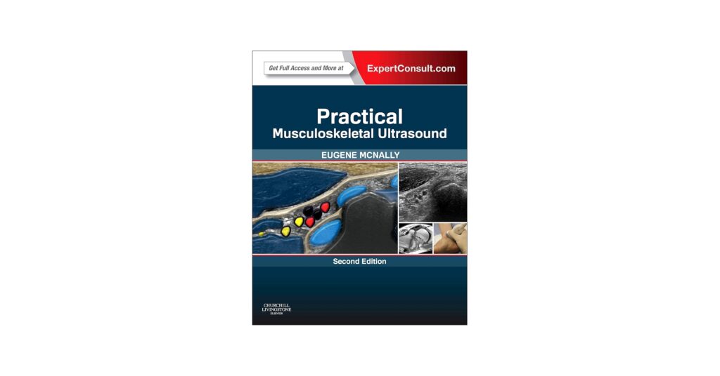
Having already acquired anatomical knowledge and a systematic approach to the work, it is necessary to have specialized books to deepen one’s understanding, study injuries, and learn to recognize them. Two of the best books for this purpose are:
- Musculoskeletal Ultrasonography, by Eugene McNally (Get the book here)
- Ultrasound of the Musculoskeletal System, by Bianchi and Martinolli
The first book is more practical and easier to read, offering numerous tips for daily practice, as well as information on common pathologies, study methodologies, and protocols; it is recommended as a first read. The second book is recommended after acquiring some knowledge and experience, and it delves a little deeper into various pathologies and their detection methods.
For any comments or suggestions: radiologyzones@gmail.com
This material was automatically translated from medicosradiologos.com.ar